Empty Bicipital Groove Mri

There were no other significant findings and there were no signs that the absence of the long head of the biceps tendon lhbt was due to a tear namely there was no intraarticular stump no abnormalities on the usual topography of the biceps anchor and no fluid in the.
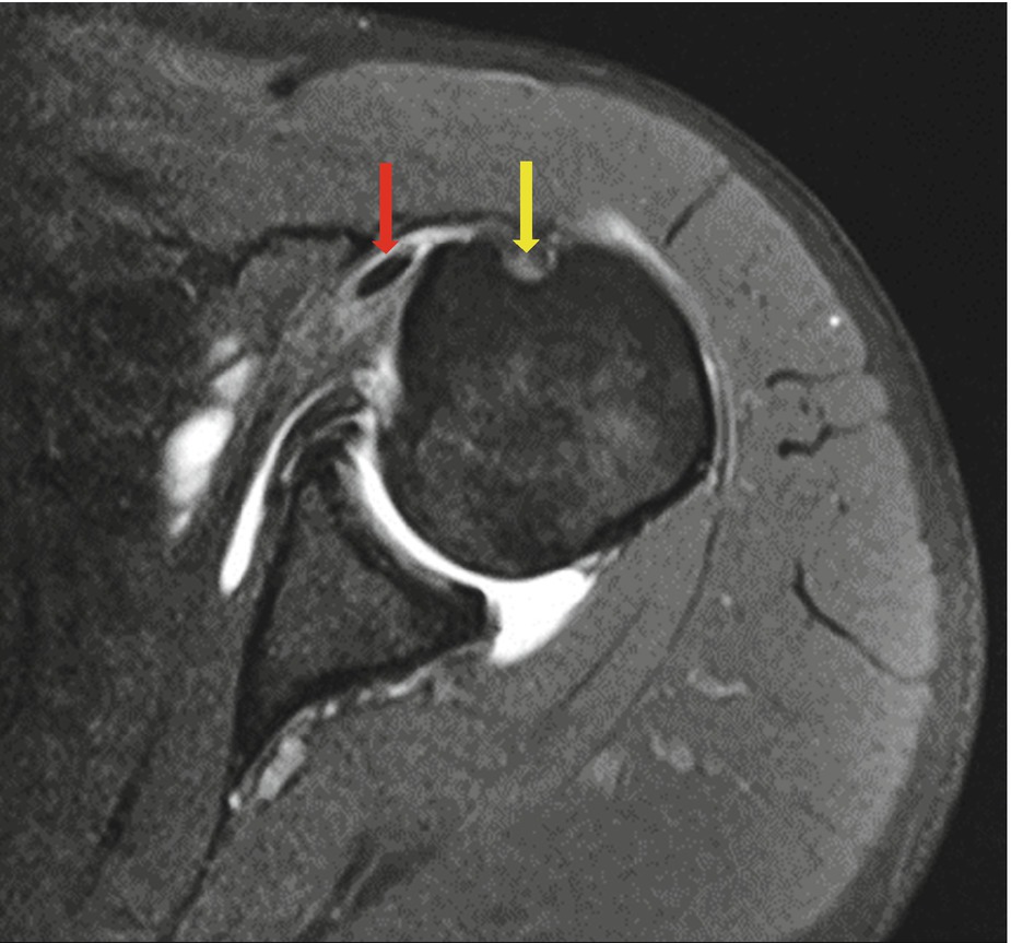
Empty bicipital groove mri. Axial pdw fat saturated mri shows high signal arrow within the tendon indicating partial thickness tear. Sirico f castaldo c baioccato v et al. If the tendon cannot be identified then a complete tear of the tendon should be sought. Red arrow shows the absence of the long biceps tendon in the pulley.
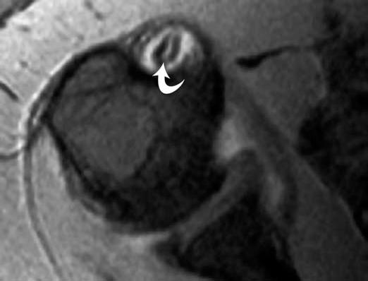
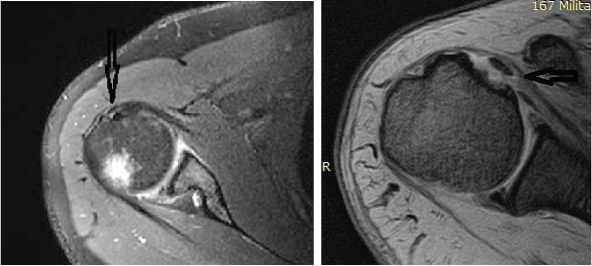
A axial pdw fat saturated mri and b short axis us scan of lhb show the absence of the tendon and empty bicipital groove arrow conflict of interest. Diagnosis is best made on axial mr images where the bicipital groove is seen to be empty and the tendon can be identified medially. It is often not possible to distinguish between type 1 and type 2 lesions at mri and in both cases the biceps tendon is often perched on the lesser tuberosity. When only the medial sheath is torn type 2 the biceps tendon is also allowed to sublux medially within the bicipital groove.
On radiographs and ct images a round lytic area in the humeral diaphysis is created when a screw is placed to anchor the distal biceps tendon to the humerus. At mri the biceps tendon is seen to be attached to the humeral shaft. C mri of the right shoulder coronal view. High t2 signal intensity noted at the of this humeral head at the biceps groove.
High t2 fluid signal intensity is noted around the thickened biceps tendon which is seen within its groove suggestive of tenosynovitis. An irregular tear of the superior glenoid labrum is also demonstrated short arrow. D mri of the right shoulder sagittal view. Thickened with intermediate high t2 signal intensity noted at the supraspinatus tendon suggestive of tendinosis.
Shoulder anatomy mri normal anatomy variants and checklist robin smithuis and henk jan van der woude. Blue arrow shows the empty intertubercular groove. A and b mri of the right shoulder axial view. The bicipital groove is empty with no biceps tendon arrowhead.
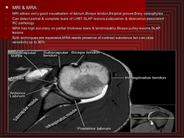
The translucent screw can be seen at mri. The fibers of the subscapularis tendon hold the biceps tendon within its groove. At this level study the middle ghl and the anterior labrum. Left shoulder mri showed an empty shallow bicipital groove with a normal robust short head of the biceps tendon shbt.
This should not be mistaken for a cyst or tumor. Mri allows for visualization of the long head of the biceps tendon. Non visualization of long head of biceps tendon in bicipital groove with medially displaced tendon. A thickened and edematous lbt arrows is identified anterior to the humerus at a level distal to the bicipital groove outlined by surrounding mild edema.


