Bicipital Groove X Ray Position

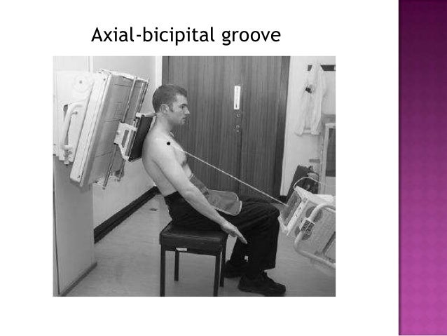
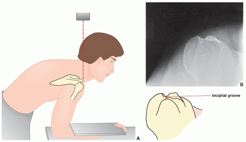
The hand is rotated 45 degrees laterally from prone to bring the bicipital groove in profile centering o the of the beam central ray is directed cranially along the long axis of humerus.
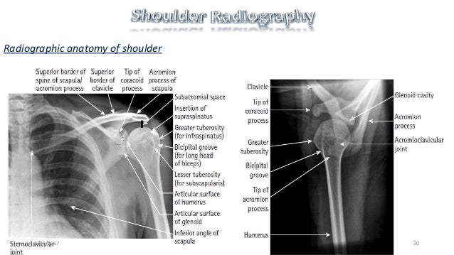
Bicipital groove x ray position. While achieving anteroposterior shoulder x ray in neutral position the patient is erect or in supine position. Central x ray should be directed to 2 5 cm inferior to the coracoid process. Ergonomic evaluation of your workstation. The tendon of the subscapularis muscle attaches both to the lesser tubercle aswell as to the greater tubercle giving support to the long head of the biceps in the bicipital groove.
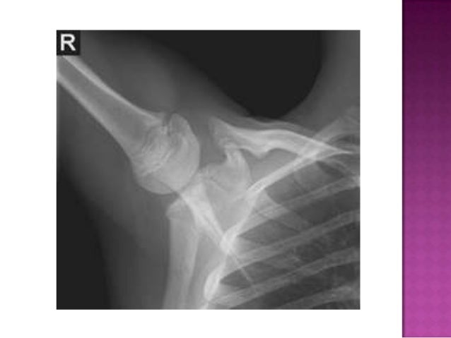
The x ray may show bony growth near the bicipital tunnel and the mri or ct would show inflammation or tearing of the tissues. This movement is one of the most specific findings of biceps tendon injury. External rotation of the arm and humeral head places the tender bicipital groove in a posterolateral position. The stryker notch view is a specialized projection of the shoulder aimed at assessing the posterior humerus.
Conservative treatment of biceps tendonitis is likely to center around reducing the damaging positions in the individual s activities of daily living. This tool allows you to search snomed ct and is designed for educational use only. Indications the stryker notch view can be used post anterior glenohumeral dislocation assessing for hill sachs lesions 1. Dislocation of the long head of the biceps will inevitably result in rupture of part of the subscapularis tendon.
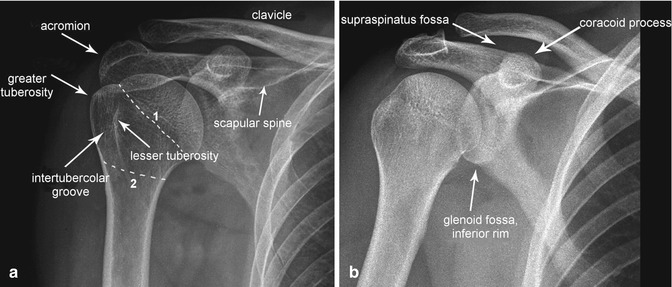
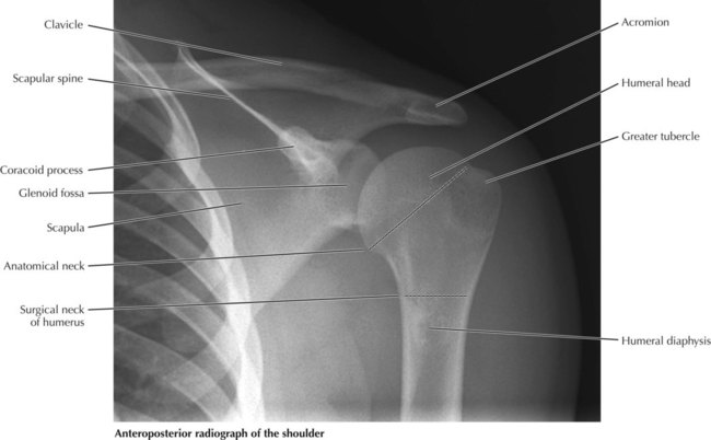
Anteroposterior shoulder view allows assessment of especially the humeral head lesions and clavicular fractures. 3 1 anteroposterior shoulder radiograph. The effect of the direction of the x ray beam on the image of the groove was studied in cadaver specimens. Patients should be changed into a hospital gown with radiopaque items removed e g.
Both shoulders and hips equidistant from the table trolley. Belts zippers the patient should be free from rotation. Bicipital groove x ray bicipital groove x ray procedure hide descriptions. The purpose of this paper was to find a standard radiographic view for measurements of the intertubercular groove of the human humeral head.
The patient is supine lying on his or her back either on the x ray table preferred or a trolley. A standardized optimal x ray beam direction was subsequently used in a clinical study on 30 patients. Shoulder tangential intertubercular bicipital groove.

