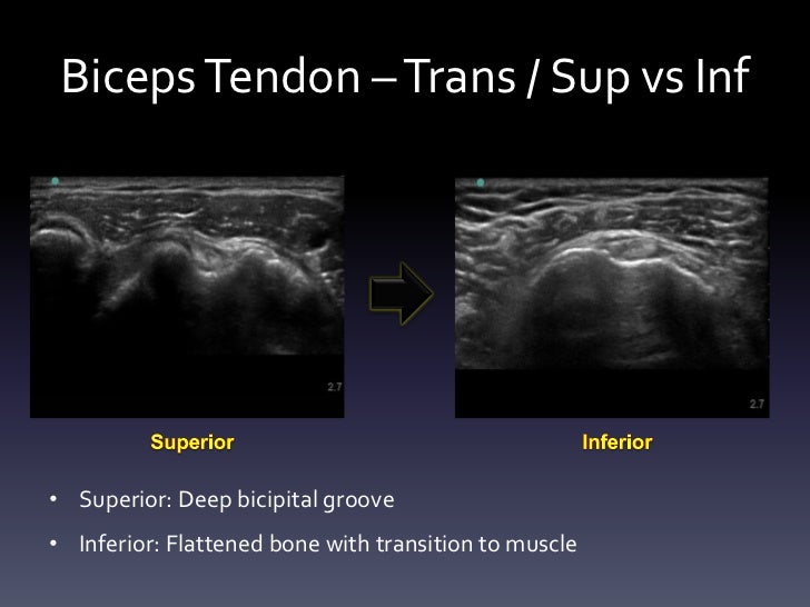
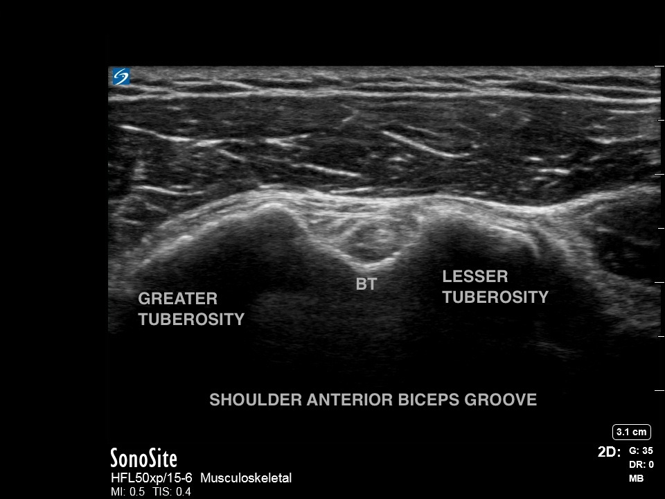
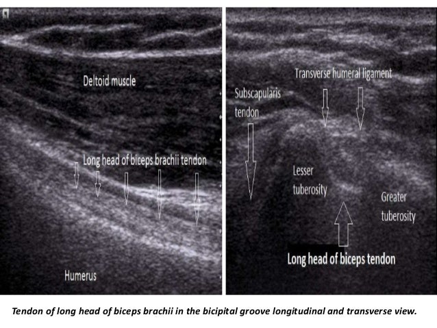
Bicipital Groove Ultrasound

Diagnosis is best made on axial mr images where the bicipital groove is seen to be empty and the tendon can be identified medially.
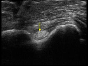
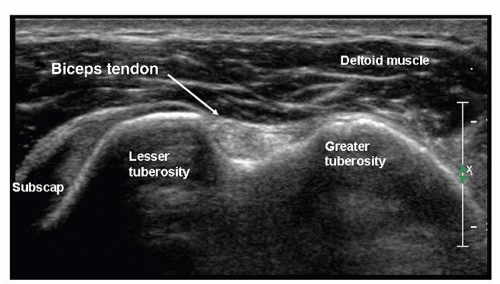
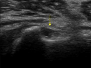
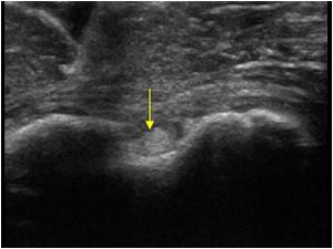
Bicipital groove ultrasound. Non visualization of long head of biceps tendon in bicipital groove with medially displaced tendon. Long head of biceps brachii tendon pathology can be examined both with ultrasound and or mri. From the gülhane military medical academy department of physical medicine and rehabilitation turkish armed forces rehabilitation center bilkent ankara turkey. The relatively poor blood supply limits the ability of these.
Inflammation of the biceps tendon in the bicipital groove which is known as primary biceps tendinitis occurs in 5 percent of patients with biceps tendinitis. A comparison of physical examinations with musculoskeletal ultrasound in the diagnosis of biceps long head tendinitis. Ultrasound images clips biceps tendon tendinosis with a thickened biceps tendon and effusion in an irregular bicipital groove. Efficacy of ultrasound in the diagnosis of biceps tendon dislocation.
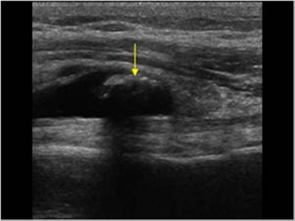
For the diagnosis of full thickness lhb tendon tear versus other findings partial thickness tear nontear abnormalities or normal in patients who underwent surgery ultrasound showed a sensitivity of 88 a specificity. Both instability and tears can result in pain and decreased function. If the tendon cannot be identified then a complete tear of the tendon should be sought. In addition ultrasound identified a small bone spur in the proximal intertubercular groove adjacent to the biceps tendon.
Signs in physical examination correlating with bicipital tendinitis are pain at the bicipital groove and a positive provocative test such as speed s test and yergason s test.
