Bicipital Groove Radiology

Long Head Of Biceps Tendon Dislocation Radiology Reference

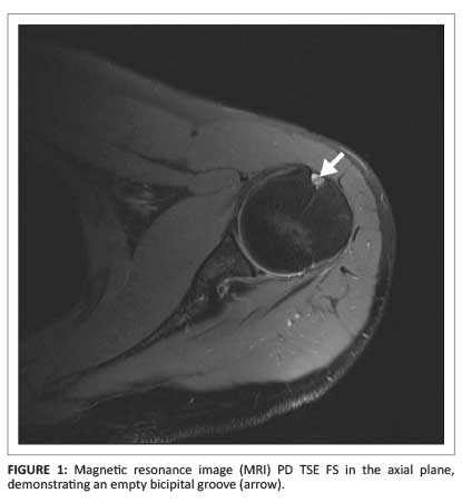
Mri Showed Normal Position Of Biceps Tendon In The Bicipital

Pathology Of The Long Head Of The Biceps Tendon Radsource

Complete Tear Of The Long Head Biceps Tendon Groove Entry

Proximal Rupture Of Long Head Biceps Brachii Tendon Radiology

Long Head Of Biceps Tendon Dislocation Radiology Reference

An accessory biceps tendon in the groove should not be mistaken for a tear.
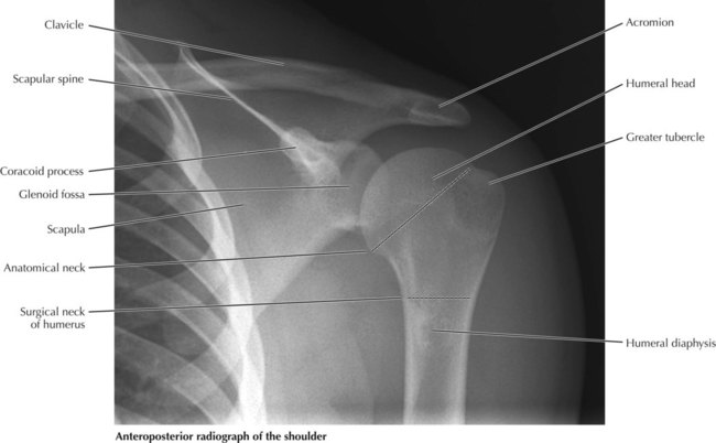
Bicipital groove radiology. The bicipital groove also known as the intertubercular sulcus or sulcus intertubercularis is the indentation between the greater and lesser tuberosities of the humerus that lodges the biceps tendon. It contains the tendon of the long head of the biceps brachii muscle which is ensheathed in a synovial reflection of the. Biceps tendon laxity in the bicipital groove may lead to superior extension of degenerative changes in the rotator interval 7 18 20. Our cases are limited to patients with a history of previous trauma to the shoulder and ensuing shoulder pain whose routine x ray studies were negative.
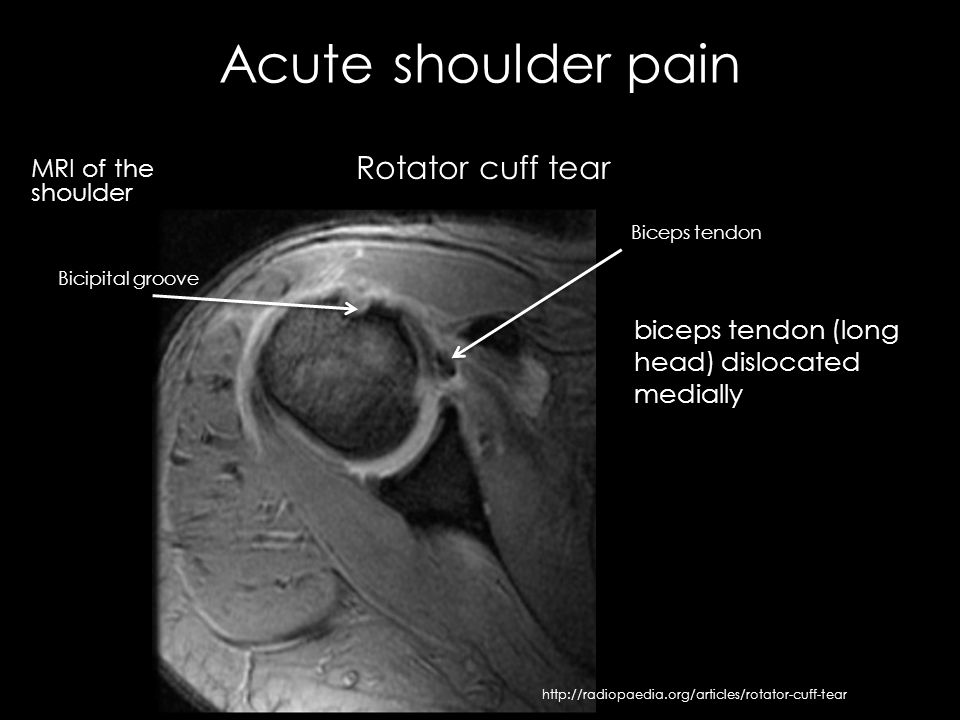
Both to the lesser tubercle aswell as to the greater tubercle giving support to the long head of the biceps in the bicipital groove. Reader 2 with 2 years of experience in musculoskeletal radiology at three levels of the bicipital groove superior middle and inferior with the shoulder in neutral position in external rotation and in internal rotation. The long head of biceps lhb tendon is usually located inferiorly in the bicipital groove held there by the biceps pulley the stabilization role of the transverse humeral ligament is controversial 3 as it moves superiorly it arches through the rotator cuff interval where it is held by a sling. The lbt was evaluated by two independent musculoskeletal radiologists f m b.
We made special tangential views on 40 such patients and in no instance was a normal groove depicted. Dislocation of the long head of biceps tendon is one of the complications of shoulder injury. Long head of biceps can be affected by numerous pathological processes 3. Pathologic changes in the bicipital groove region.
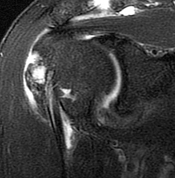
The dislocated tendon can be identified medial to the bicipital groove best seen on the axial and oblique coronal and sagittal images. High t2 signal intensity noted at the of this humeral head at the biceps groove. The accessory biceps tendon often originates from the supraspinatus tendon. Thickened with intermediate high t2 signal intensity noted at the supraspinatus tendon suggestive of tendinosis.
The magnetic resonance mr images from six patients with biceps tendon dislocation two in whom it was surgically proved and four in whom it was suspected were retrospectively evaluated. Bencardino j presented at the 2011 annual meeting of the society of skeletal radiology and the 2012 arrs annual meeting. High t2 fluid signal intensity is noted around the thickened biceps tendon which is seen within its groove suggestive of tenosynovitis. Potential causes of pulley lesions include a congenital rotator interval defect 23 a supratubercular ridge as an osseous protrusion of the lesser tuberosity 24 or a shallow groove 5.
Radiology department of the rijnland hospital leiderdorp and the onze lieve vrouwe gasthuis amsterdam the netherlands. The bicipital groove is typically 4 6 mm deep 1.
Long Head Of The Biceps Tear A Mri Arrow Bicipital Groove With

A Profile View Of A Humeral Head Segmented From A Mri Of The
Pathology Of The Long Head Of The Biceps Tendon Radsource

Images Of Note 3t Mri Reveals Biceps Tear With Intra Articular
Complete Rupture Of The Long Head Of The Biceps Tendon And The
Long Head Of The Biceps Tear A Mri Arrow Bicipital Groove With

Tenosynovitis Of Biceps Tendon Radiology Case Radiopaedia Org

Vessels Adjacent To The Long Head Of The Biceps Tendon Arrows
Proximal Rupture Of Long Head Biceps Brachii Tendon Radiology

The Radiology Assistant Shoulder Mr Anatomy
Upper Limbs Radiology Key
Msk Clinical Cases Traumatology Ppt Video Online Download
The Radiology Assistant Shoulder Anatomy Mri
